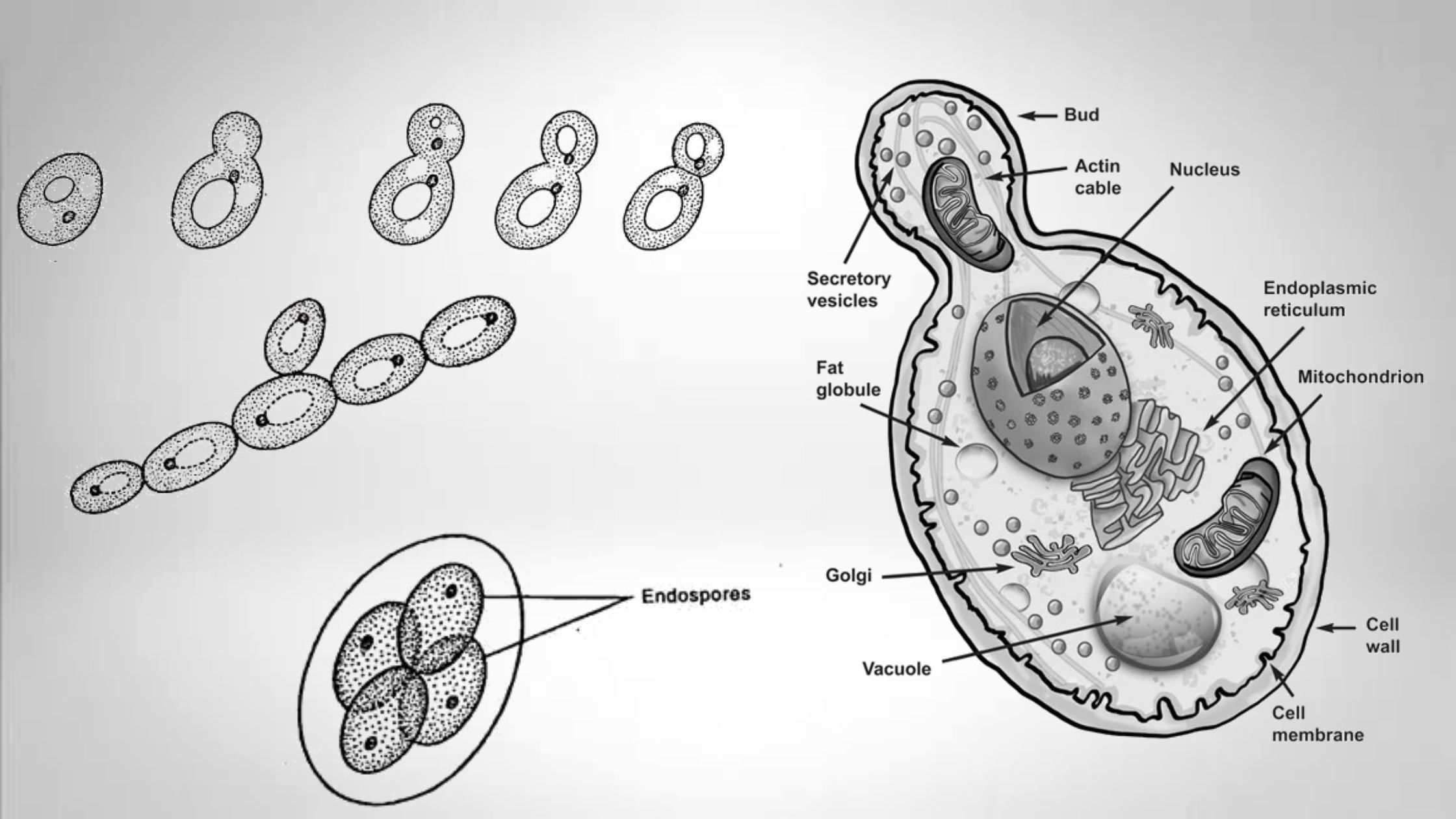
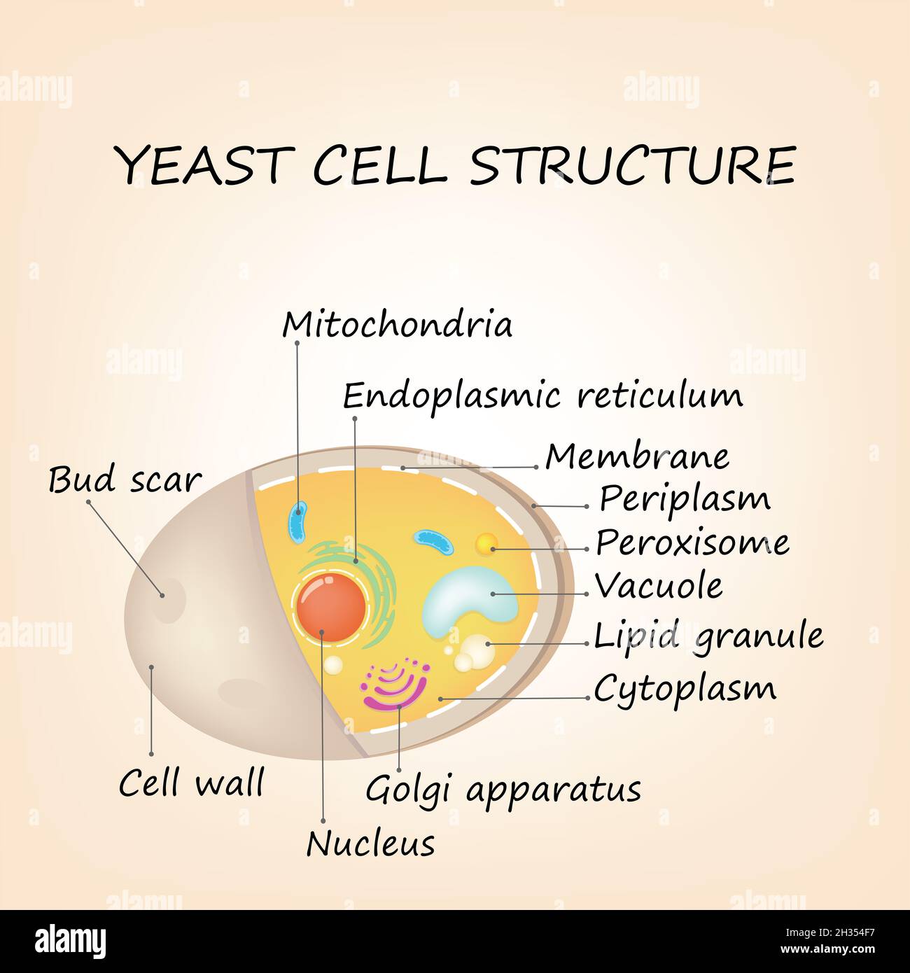
Saccharomyces Cerevisiae Microscope Labeled
Dec. 21, 2024
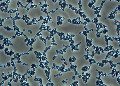
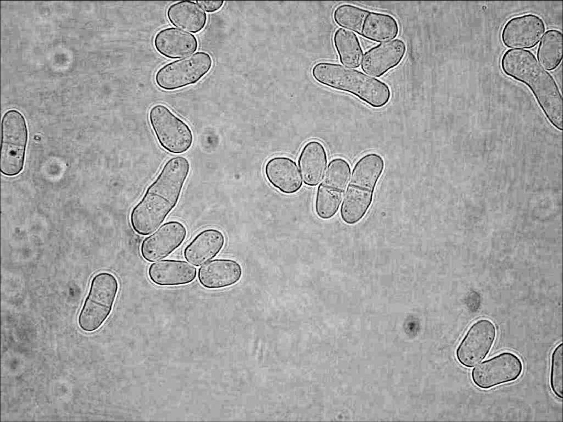
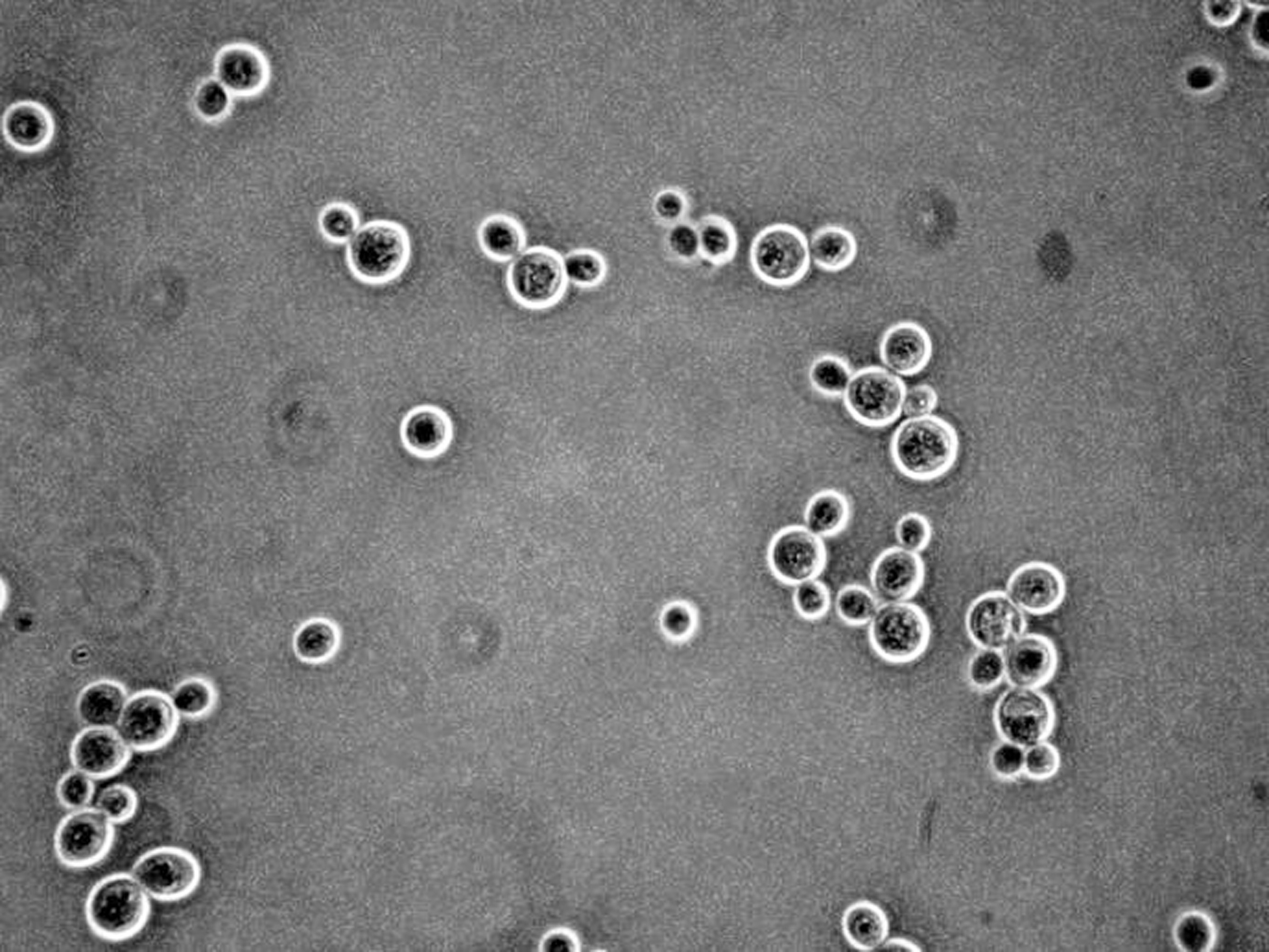
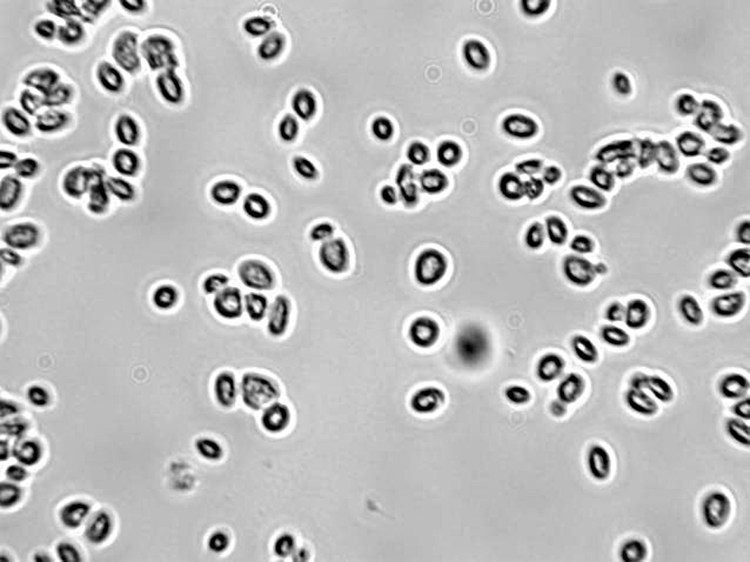
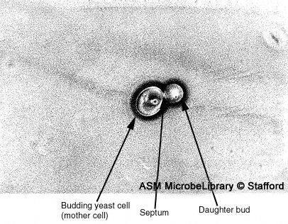
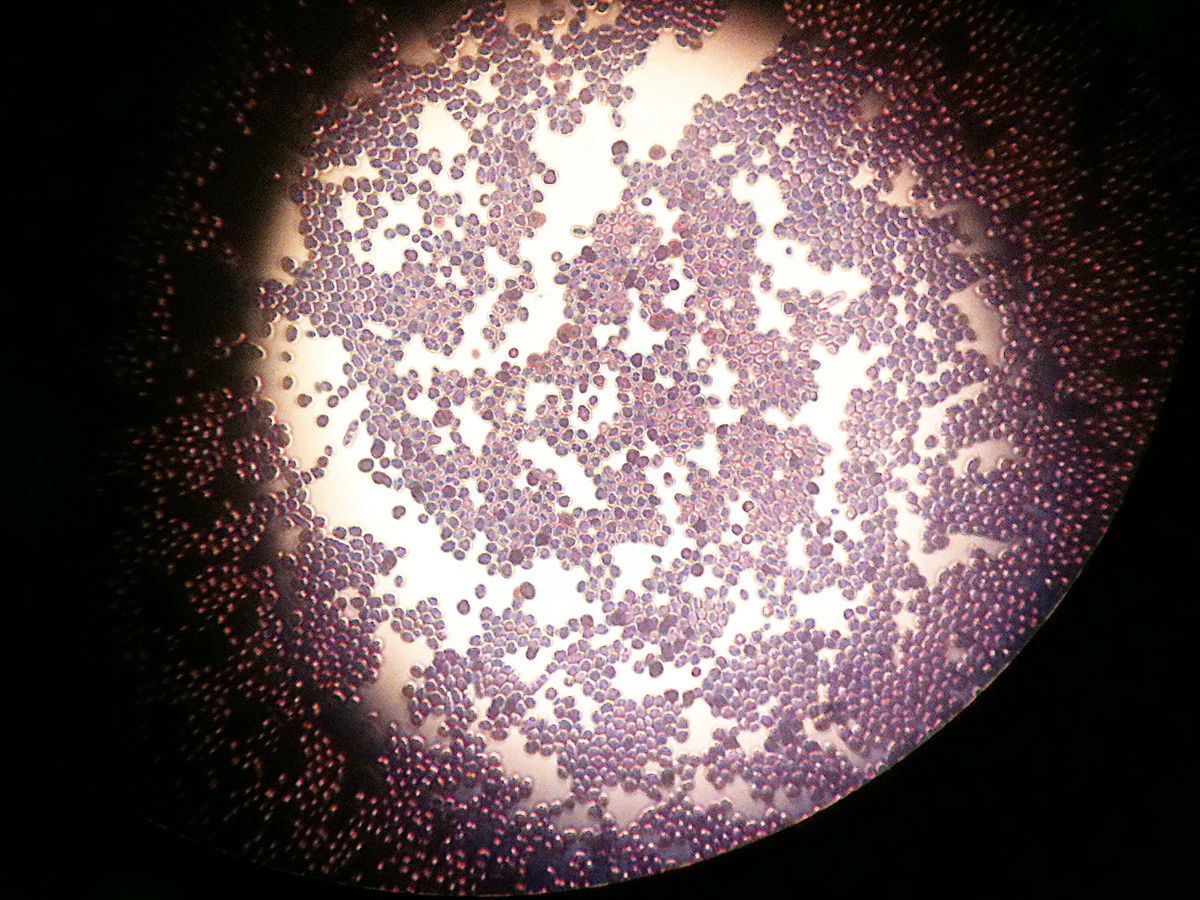
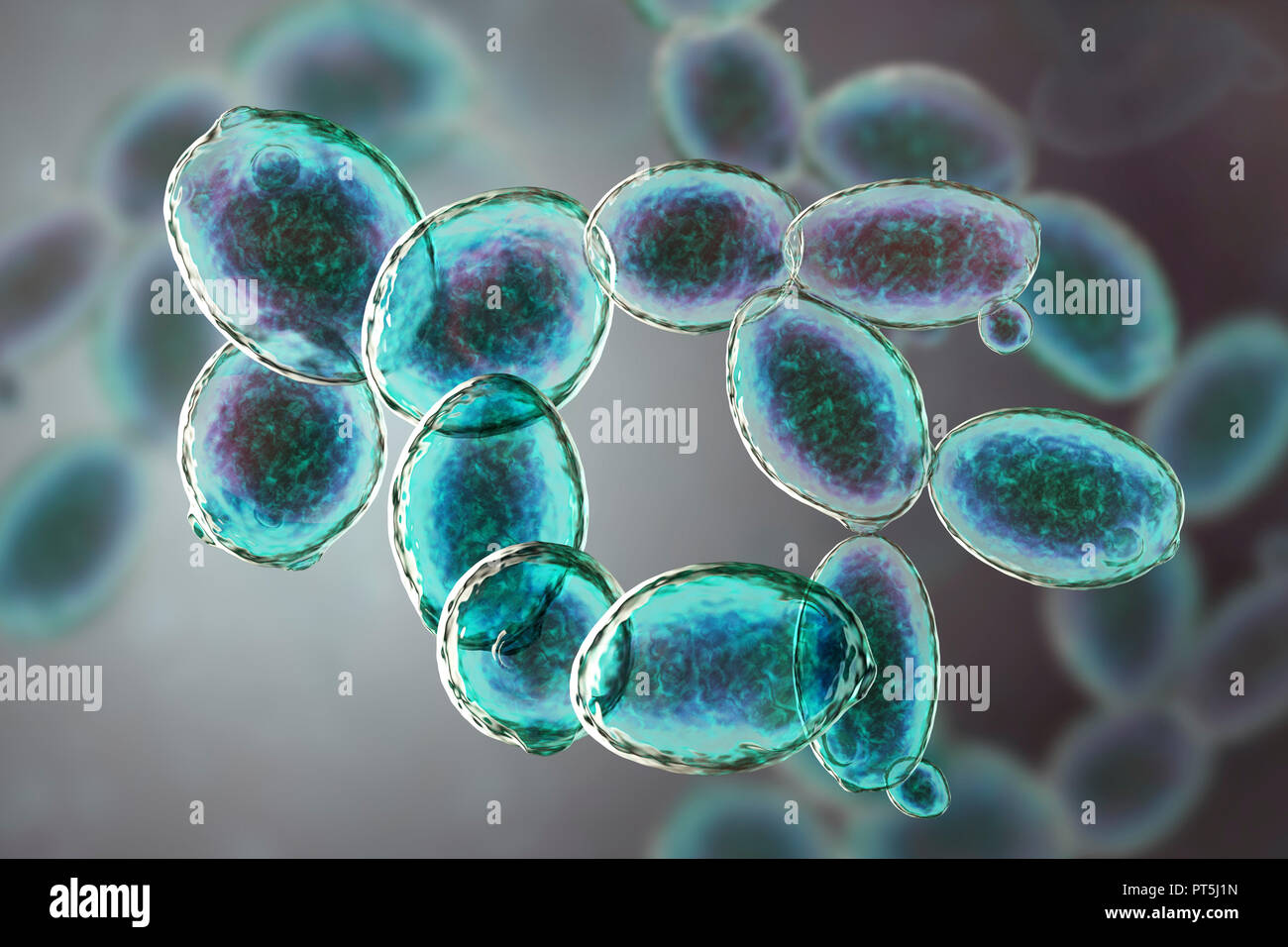
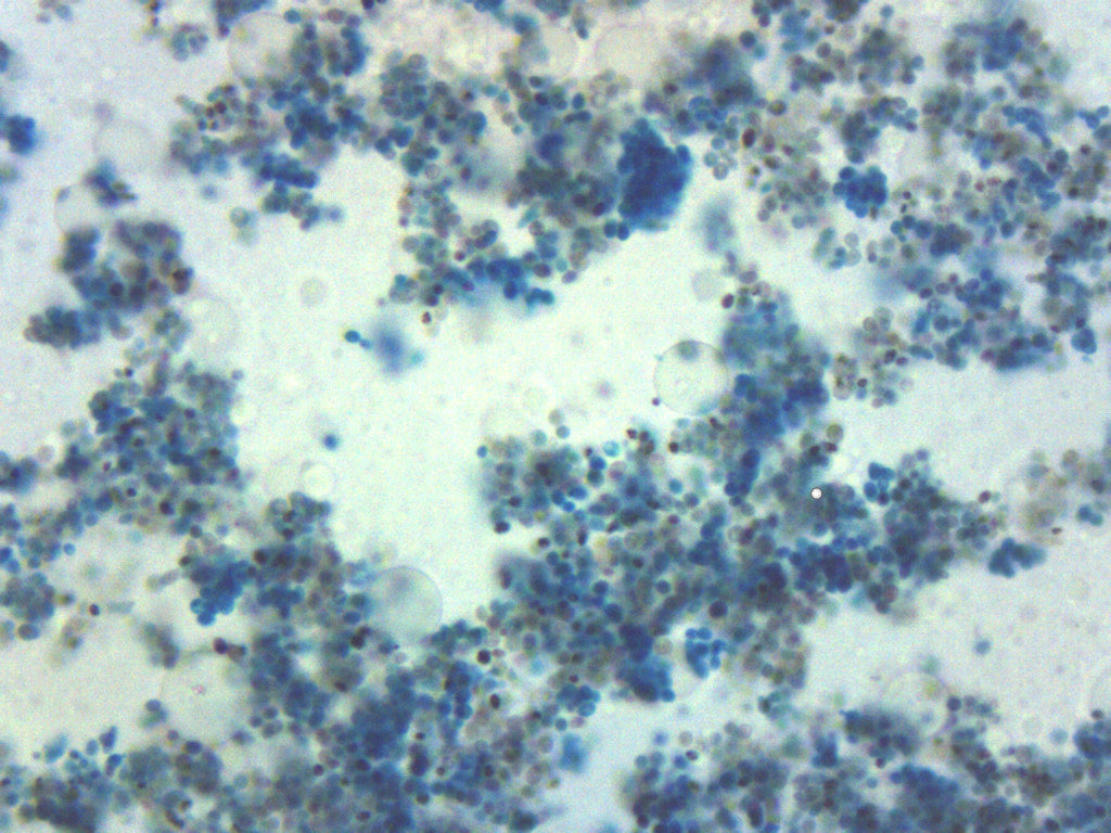
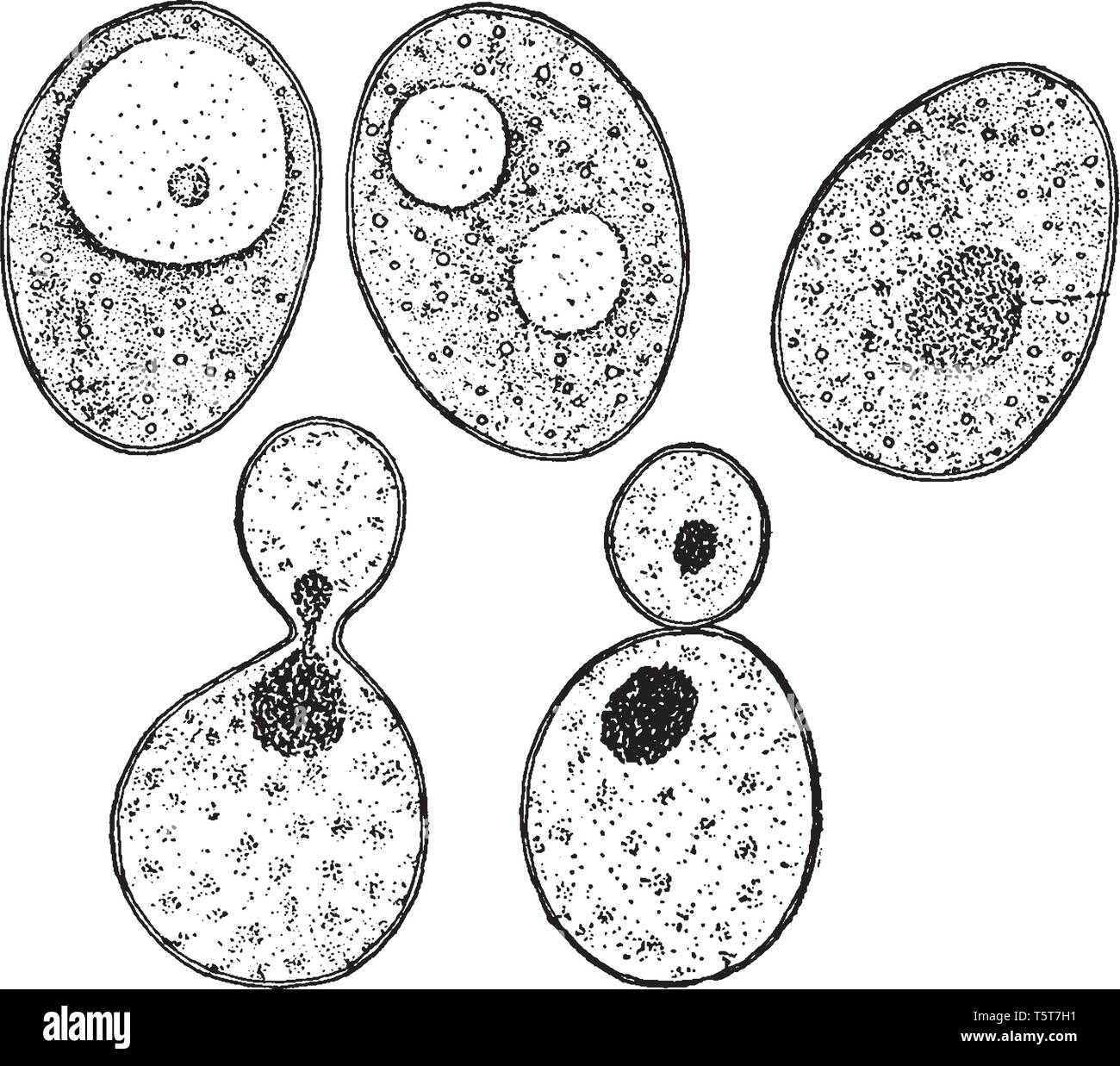
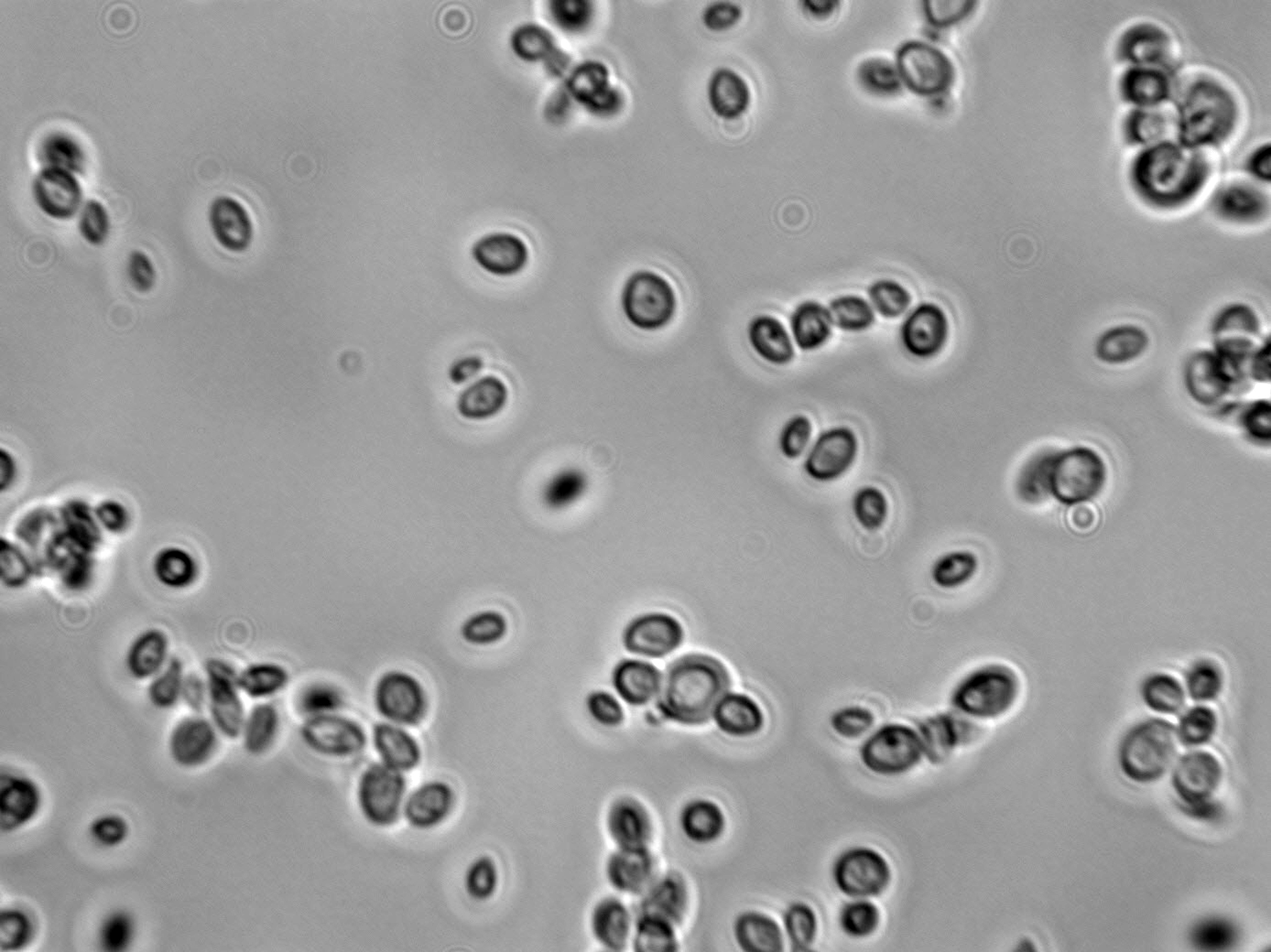
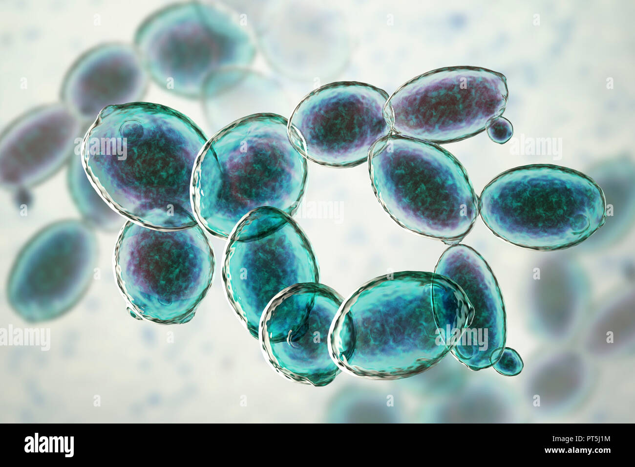
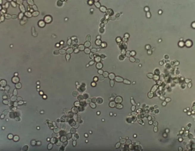
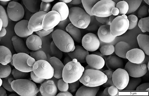
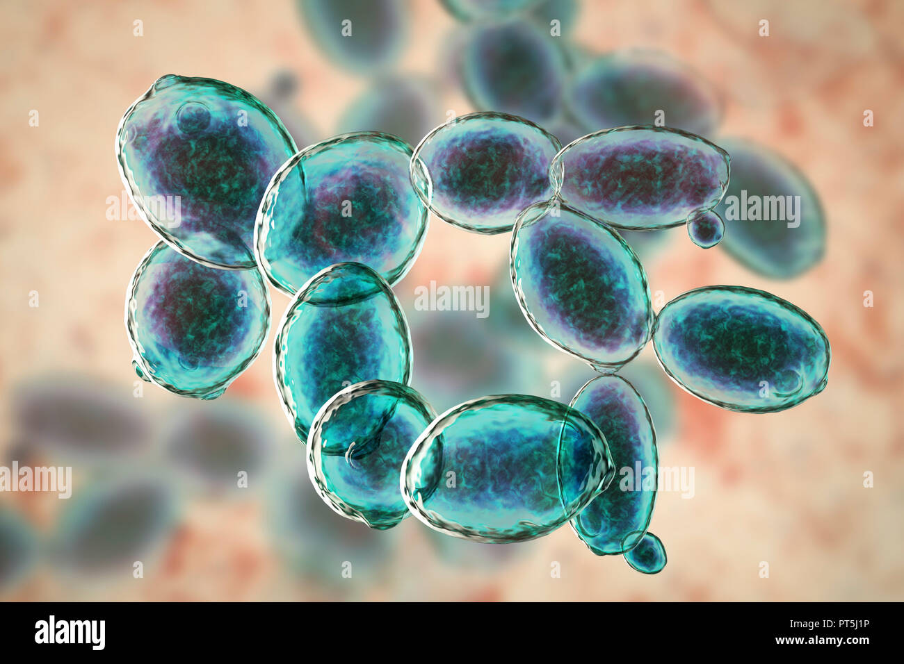
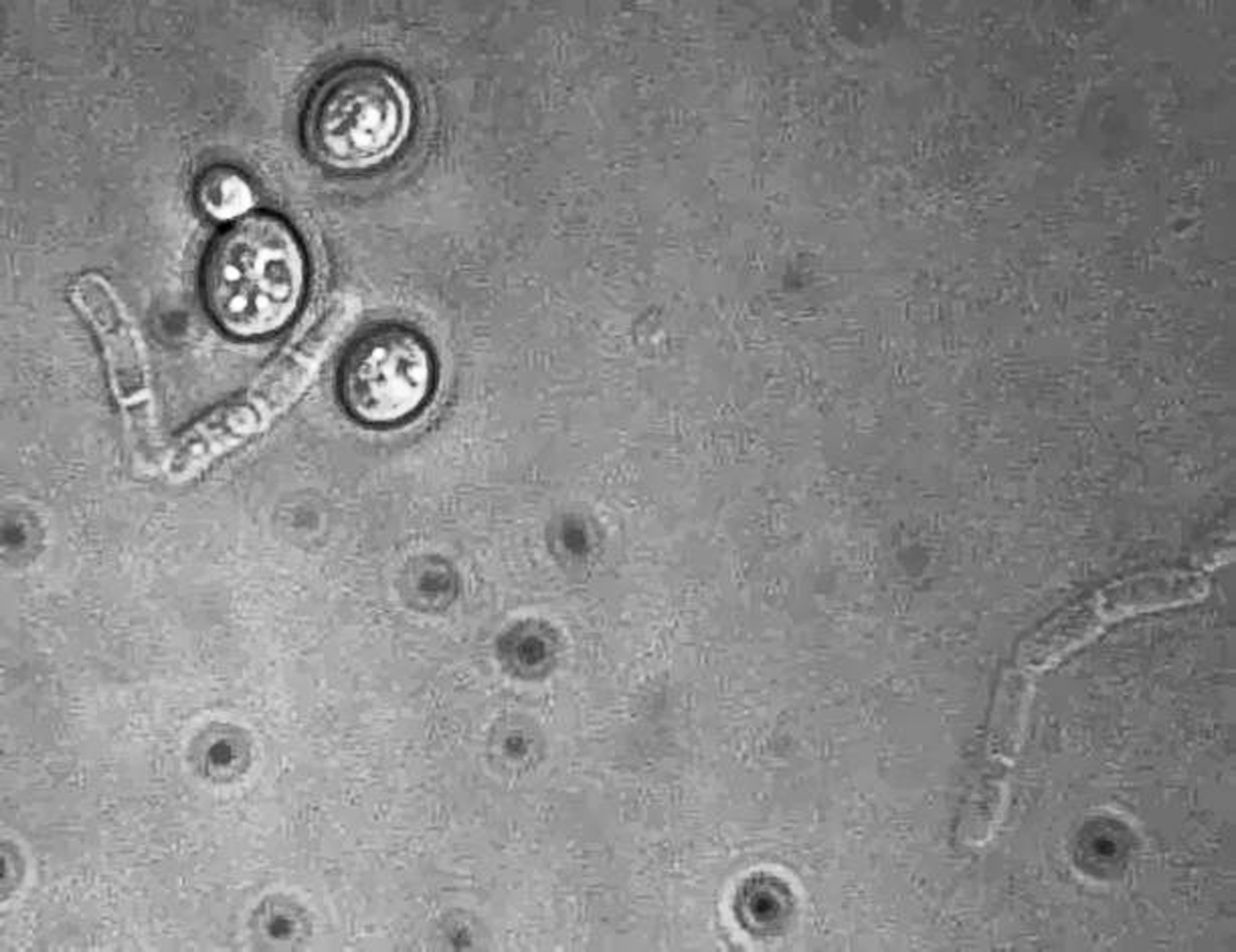
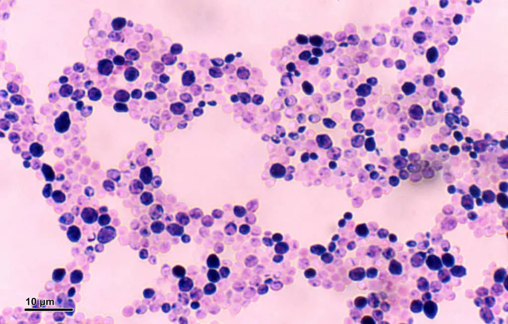
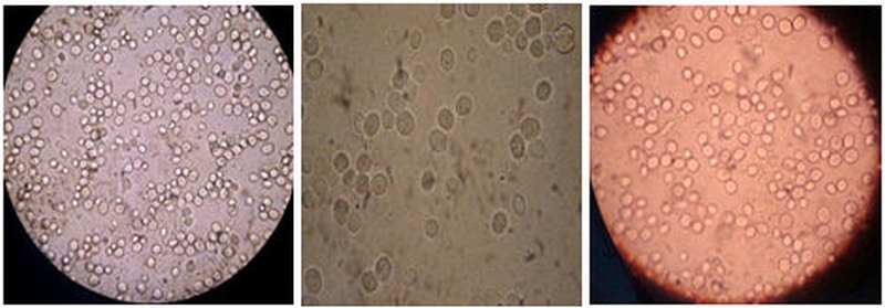
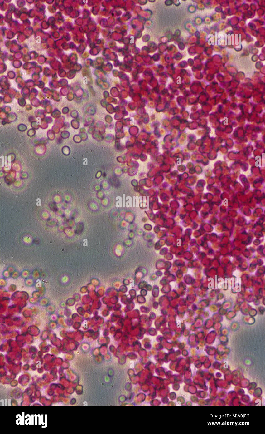
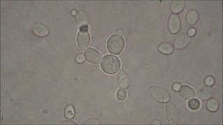
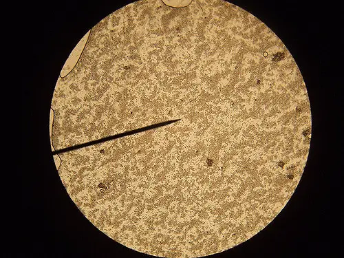
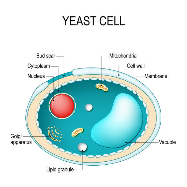
Light microscopy image of Saccharomyces cerevisiae in pH=4 and DO5% | Download Scientific Diagram microscopic identification of S. cerevisiae. The typical budding... | Download Scientific Diagram − Microscopic image of waste Saccharomyces cerevisiae [31]. | Download Scientific Diagram 2: Yeast cell; a) colonies of S. cerevisiae on agar plate (www, 2005),... | Download Scientific Diagram Scanning Electron Microscope Blog: Fungi - Images for Eastfield College Microbiology File:Saccharomyces cerevisiae 100x phase-contrast microscopy.jpg - Wikimedia Commons | Saccharomyces Cerevisiae Microscope Labeled
![− Microscopic image of waste Saccharomyces cerevisiae [31]. | Download Scientific D…](https://www.researchgate.net/publication/314207518/figure/fig2/AS:467889854652417@1488565042808/Microscopic-image-of-waste-Saccharomyces-cerevisiae-31.png)
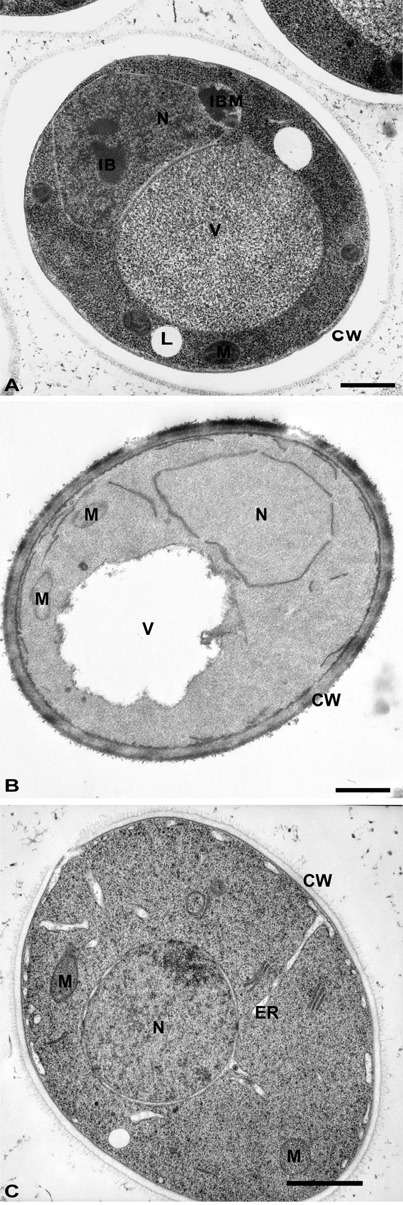
![PDF] 2015A Frankl Microbial Cell | Semantic Scholar](https://d3i71xaburhd42.cloudfront.net/2255eeb4fd9f36aed7794b26f532a3d947202172/7-Figure2-1.png)